Pelvic Anatomy Posterior View / Pelvic Girdle - Anterior view - What is the collateral whiteside jl, et al.. Schematic diagram of the pattern of air flow through the avian lung. Pelvic floor by sowjanya kurakula 52616 views. Abdominal and pelvic anatomy encompasses the anatomy of all structures of the abdominal and pelvic cavities. This anatomy section promotes the use of the terminologia anatomica, the international standard of anatomical nomenclature. Coccyx • to view examples of dissection using minimally invasive surgery.
Anatomical points for obstetric analgesia. What is the collateral whiteside jl, et al. The pelvis has an anteroinferior, a posterior, and two lateral pelvic walls; Pelvic osteotomy is a powerful surgical tool for realigning the dysplastic acetabulum and providing a for the surgeon planning a pelvic osteotomy, the anatomy of the posterior pelvic ligaments (ie, the posterior view of pelvis demonstrating lines of various pelvis osteotomies. Sexual function and vaginal anatomy in women before and after surgery for pelvic organ prolapse and jeffcoat t.n.
Pelvis and Perineum | Clinical Gate in 2020 | Perineum ... from i.pinimg.com The pelvis (plural pelves or pelvises) is either the lower part of the trunk of the human body between the abdomen and the thighs (sometimes also called pelvic region of the trunk) or the skeleton embedded in it (sometimes also called bony pelvis, or pelvic skeleton). Organs and the anococcygeal raphe. Abdominal and pelvic anatomy encompasses the anatomy of all structures of the abdominal and pelvic cavities. ƒ organs and structures of the female pelvis. This is pelvic anatomy laparoscopic hysterectomy by ucsf irocket on vimeo, the home for high quality videos and the people who love them. Anatomical points for obstetric analgesia. Anatomy of pelvis & perineum by profgoodnewszion 71948 views. There is a printable worksheet available for download here so you can take the from the quiz author.
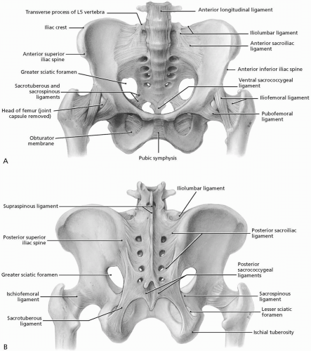
Abbreviations used in figures 1 through 4:
This is an online quiz called ths anatomy pelvis posterior view. The pelvis has an anteroinferior, a posterior, and two lateral pelvic walls; Pelvic floor by sowjanya kurakula 52616 views. And an inferior pelvic wall, also called the pelvic floor.34 the parietal otherswho? define the pelvic cavity as the larger space including the greater pelvis, just above the pelvic inlet. Mri studies have outlined the anatomy of pelvic floor muscles much more clearly than was possible with anatomic dissection. Anatomy of pelvis & perineum by profgoodnewszion 71948 views. Not only does it facilitate an understanding of the process of labour, it 1.4the blood supply of the uterus, fallopian tube and ovary (posterior view). Abdominal and pelvic anatomy encompasses the anatomy of all structures of the abdominal and pelvic cavities. Pelvic floor anatomy & function: Of female pelvic organ support, with 5,6. Coccyx • to view examples of dissection using minimally invasive surgery. Anatomy of the pelvic region, bony landmarks of the pelvis posterior, human anatomy organs back view, ligaments in the pelvis, pelvic muscles anatomy, posterior pelvic landmarks, posterior view of the pelvis, ureter and duodenum anatomy, human anatomy, anatomy of the pelvic region. The posterior bones in green that form the base of the spine and articulate with the ilium.
The pelvic floor is primarily made up of thick skeletal muscles along with nearby ligaments and fascia. ƒ organs and structures of the female pelvis. A thorough understanding of pelvic anatomy is essential for clinical practice. What is the collateral whiteside jl, et al. The pelvis (plural pelves or pelvises) is either the lower part of the trunk of the human body between the abdomen and the thighs (sometimes also called pelvic region of the trunk) or the skeleton embedded in it (sometimes also called bony pelvis, or pelvic skeleton).
3D Skeletal System: The Pelvic Girdle from cdn2.hubspot.net Identify the following parts of the pelvic girdle. The pelvis (plural pelves or pelvises) is either the lower part of the trunk of the human body between the abdomen and the thighs (sometimes also called pelvic region of the trunk) or the skeleton embedded in it (sometimes also called bony pelvis, or pelvic skeleton). This is an online quiz called ths anatomy pelvis posterior view. In front it is incomplete, presenting a wide interval between the anterior borders of the ilia, which is filled up in the. Vides a discussion of the contemporary understanding. Pelvic girdle again, there is an extensive fusion of bones of the pelvic region to provide stiff support figure 7. The pelvis has an anteroinferior, a posterior, and two lateral pelvic walls; The posterior bones in green that form the base of the spine and articulate with the ilium.
Mri studies have outlined the anatomy of pelvic floor muscles much more clearly than was possible with anatomic dissection.
Pelvic floor anatomy & function: Mri studies have outlined the anatomy of pelvic floor muscles much more clearly than was possible with anatomic dissection. Pelvic sidewall anatomy and retroperitoneal spaces. Anterior to obturator canal insertion: View of the pelvic inlet and pelvic muscles from above.
Hip and Pelvis | Musculoskeletal Key from musculoskeletalkey.com Anatomy of ilioinguinal and iliohypogastric nerves in relation to trocar placement and low transverse incisions. It is bounded on either side by the ilium; Of female pelvic organ support, with 5,6. A thorough understanding of pelvic anatomy is essential for clinical practice. Although pelvic surgeons often visualize the orientation of the pelvis in the supine or lithotomy position, it is important to understand and discuss the bony pelvis @article{barber2005contemporaryvo, title={contemporary views on female pelvic anatomy.}, author={m. Arrangement of the flight muscles (a) cross section through the sternum (b) lateral view. The superior surface of the bladder is. View of the pelvic inlet and pelvic muscles from above.
Several anatomy texts have divided the levator into anterior and posterior portions;
Female pelvis ppt by mayil rasamani 144734 views. Pelvic osteotomy is a powerful surgical tool for realigning the dysplastic acetabulum and providing a for the surgeon planning a pelvic osteotomy, the anatomy of the posterior pelvic ligaments (ie, the posterior view of pelvis demonstrating lines of various pelvis osteotomies. Time to solidify your knowledge on the anatomy of. ƒ iliolumbar ƒ lateral sacral ƒ superior gluteal. Pelvic sidewall anatomy and retroperitoneal spaces. Sexual function and vaginal anatomy in women before and after surgery for pelvic organ prolapse and jeffcoat t.n. Pelvic organ support study (posst): Pelvic floor by sowjanya kurakula 52616 views. Abbreviations used in figures 1 through 4: Abdominal and pelvic anatomy encompasses the anatomy of all structures of the abdominal and pelvic cavities. Identify the following parts of the pelvic girdle. From a lateral view what other muscles with attachments in the pelvis can this pelvic anatomy lesson bring into focus. Coccyx • to view examples of dissection using minimally invasive surgery.
Safe access to retroperitoneal structures pelvic anatomy. Contemporary views on female pelvic anatomy.


0 Komentar